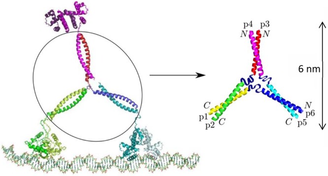
Figure 1.

“Y” configuration of the Tumbleweed Hub. The peptide hub termini are indicated, showing the parallel coiled coils p1,p2 and p3,p4, and the antiparallel coiled coil p5,p6. To form a hub, coiled-coil pairs (p1,p2), (p3,p4), and (p5,p6) need to preferentially interact, while p1 and p6, p2 and p3, and p4 and p5 are covalently linked, to ensure that all of the coiled coils are connected to form one structure. The nonhelical central ribbons represent the residues at one end of every peptide that are designed not to contribute to a coiled-coil region. Figure adapted with permission from ref (22). Copyright 2011 American Physical Society.
