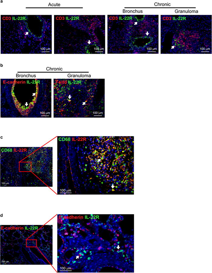
Figure 3. Epithelial cells and macrophages express IL-22R during Mtb infection.

B6 mice were aerosol infected with ∼500 CFU Mtb HN878 and lungs were harvested following acute (day 20 and 50 p.i.) or chronic (day 80 and 100 p.i.) stages of infection and formalin-fixed and paraffin-embedded (FFPE). Serial sections from FFPE sections were processed for immunofluorescence using antibodies specific for (a) CD3 (red) and IL-22R (green), (b) E-cadherin (red) or F4/80 (red) and IL-22R (green). (c) FFPE lung sections from active TB patients were processed for immunofluorescence using antibodies specific for CD68 (green) and IL-22R (red) and (d) E-cadherin (red) and IL-22R (green). All sections were counterstained with DAPI (blue). White arrows indicate cells that co-localize expression of IL-22R with CD68 or E-cadherin. n = 4-6. Original magnification for (a,b) 200×; (c,d left) 4×4 200× mosaic; (c,d right) 200×. One representative image shown.
