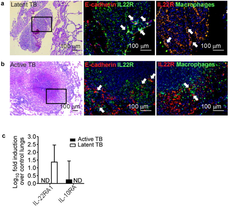
Figure 4. Preferential accumulation of IL-22+ macrophages within granulomas of latent TB.

NHPs were aerosol infected with Mtb CDC1551 and lungs were harvested from NHPs with (a) latent and (b) acute TB. FFPE lung sections were prepared and stained for (left) H&E or immunofluorescence with antibodies specific for (middle) E-cadherin (red) and IL-22R (green), or (right) macrophage (green) and IL-22R (red). All sections were counterstained with DAPI (blue). White arrows indicate co-localization of IL-22R with macrophage or epithelial markers. Original magnification for (left) H& E panel is 100×, (middle & right) immunofluorescence panels 200×. (c) Induction of IL-22RA1 and IL-10RA was measured by real-time PCR and fold induction in either latently or actively infected NHP lungs over levels expressed in control lungs is shown. n = 4-6. Error bars represent means ± s.e.m. ND, not detected.
