Fig. 3.

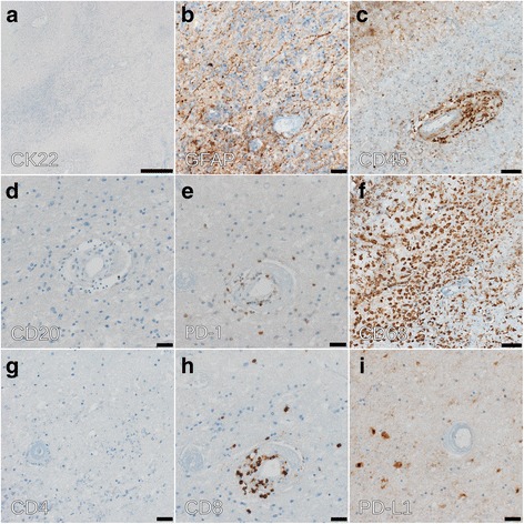
Immunohistochemical analysis of brain lesion. a Staining with CK22 monoclonal anti-cytokeratin antibody cocktail. b Staining with the astroglial marker anti-GFAP (glial fibrillary acidic protein). Scale bar: 20 μm. c CD45 (leukocyte antigen) staining for the detection of immune cells demonstrates perivascular infiltrates. Scale bar: 50 μm. d Very few CD20-positive B cells are detected. Scale bar: 20 μm. e The perivascular infiltrate are predominantly PD-1 positive T cells. Scale bar: 20 μm. f CD68 staining of macrophages highlights resorptive changes. Scale bar: 50 μm. (g, h) The perivascular T cell infiltrates are mainly CD8 T cells. Scale bar: 20 μm. i Only little PD-L1 staining was detected. Scale bar: 20 μm
