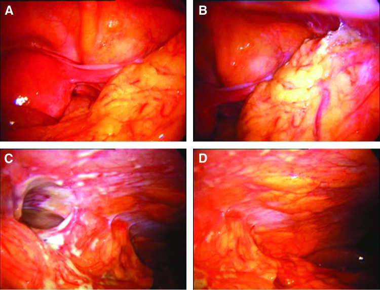
FIG. 16.
(A) Second-look laparoscopy 3 days after nonproblematic myoma enucleation of the uterine back wall. The patient experienced bouts of high temperature and had increasing inflammation parameters. No drain was inserted. The overview shows normal pelvic anatomy and no sign of peritonitis or organ damage. (B) Atypical adhesions in the right lower abdomen (cecal region). (C) The endoscope clearly demarcates the bowel damage and signs of local peritonitis. The bowel injury probably occurred when the bowel was moved out of the operating field. (D) The upper right quadrant is also free of any signs of peritonitis.

