Fig. 1.

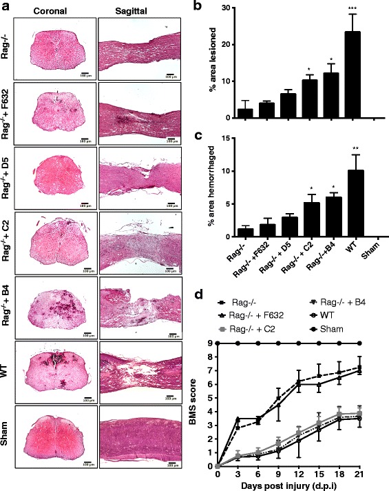
Specific IgM mAbs restore injury in otherwise protected Rag1−/− mice. a Representative H&E stained coronal and sagittal sections of WT mice, Rag1−/− mice, and Rag1−/− mice reconstituted with indicated mAb. As quantified in subsequent panels, histopathology revealed Rag1−/− mice were protected from injury, B4 and C2 mAbs restored injury in Rag1−/− mice, and control F632 and D5 mAbs had no significant effect on injury in Rag1−/− mice. b Lesion size determined from step sections obtained across 1-mm distance from the epicenter and expressed as percentage of total area of each section analyzed. Mean ± SE, ***p < 0.001, **p < 0.01, *p < 0.05 represent comparisons to Rag1−/−, n = 6 per group. c Hemorrhaged area determined from step sections obtained across 1-mm distance from the epicenter and expressed as percentage of total area of each section analyzed, Mean ± SE, ** p < 0.01, *p < 0.05 represent comparisons to Rag1−/−, n = 6 per group. d Open field 10-point BMS score to assess functional recovery over a 21-day period after SCI. A score of 9 represents no impairment of locomotor activity. Groups consisted of WTC57/bl6 and Rag1−/− mice, and Rag1−/− mice reconstituted with indicated IgM mAb. Mean ± SE, *p < 0.05, n = 6–8. Starting at day 3, significant differences were seen between Rag1−/− mice and Rag1−/− mice treated with control mAb vs. WT mice and Rag1−/− mice reconstituted with B4 and C2 mAbs
