
Fig. 2.
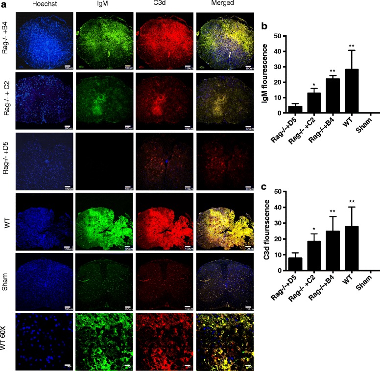
IgM binding and C3 deposition in injured spinal cords of wt and B4 mAb reconstituted Rag1−/− mice. Coronal step sections were obtained up to 0.5 mm each side from epicenter of contusion injury, 72 h after SCI. a Analysis of IgM and C3d deposition in spinal cords isolated from wt mice or Rag1−/− mice reconstituted with either B4 mAb or D5 mAb (control). Composite image (merged) indicates co-localization. Bottom panel shows high mag images of injured WT spinal cord. b, c Quantification of IgM binding and C3d deposition was done by measuring intensity from step sections across the length of 1 mm, centered at the epicenter and starting at 100 μm from the epicenter on each side. Mean ± SE, *p < 0.05, **p < 0.01, n = 6 per group. Scale bar = 100 μm