
Fig. 4.
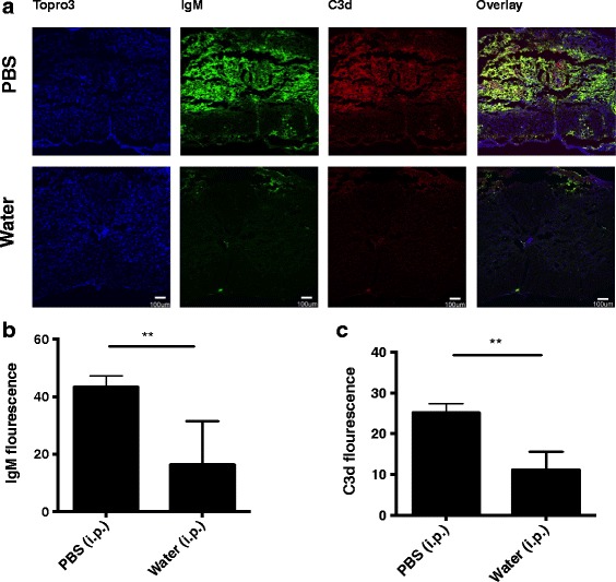
B1 B cell depletion reduced IgM deposition and complement activation in the injured spinal cord. a Immunofluorescence images of IgM and C3d deposition in cords isolated from control and B1 B cell depleted mice. Coronal step sections (100 um) were obtained across 1 mm from epicenter of contusion injury, and intensity measured and averaged. Composite image (merged) indicates co-localization. Representative images, n = 6. b, c Quantification of IgM binding and C3d deposition, respectively. Mean ± SE, **p < 0.01, n = 6