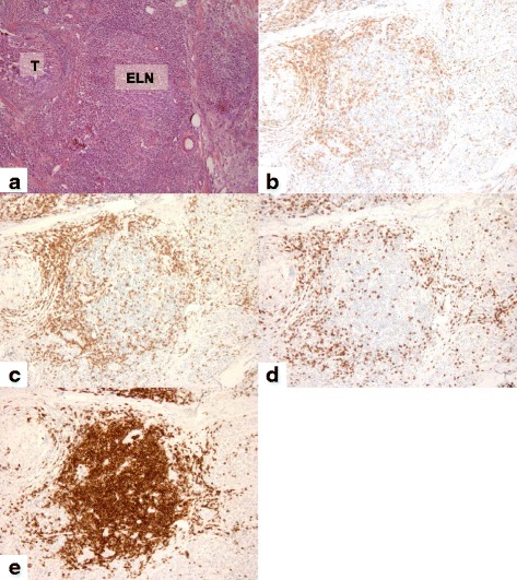
Fig. 4.

Analysis of primary breast tumors with H&E staining and IHC. Representative high-chemokine-scored breast slide revealed a marked peritumoral lymphocytic host response, organized as ectopic lymph node-like structures (ELN) by H&E staining (a) and by IHC (b–e). The immunohistochemical analysis of the lymphoid population highlighted the CD3+, CD4+, and CD8+ T cells (b, c and d) distributed in the parafollicular cortex or marginal zones and with some dispersion into the follicles while CD20 + B cells are concentrated in the center of the follicles (e). Magnification × 200
