Fig. 4.

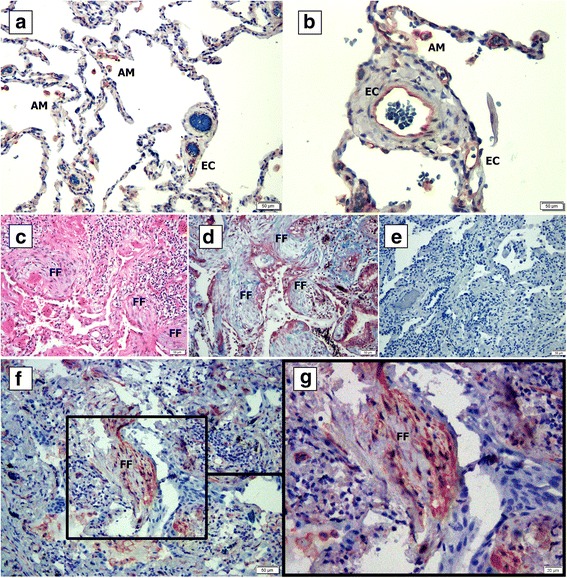
IPF fibroblastic foci express Integrin a5 (ITGA5). Normal (a-b) and IPF (c-g) lung tissue samples were fixed in formaldehyde and paraffin embedded. Following deparaffinization, slides were stained with hematoxylin and eosin (c), Masson-trichrome stain (d), IgG rabbit isotype control (e) and with anti-ITGA5 antibody (a-b, f-g). FF = fibroblastic foci; AM- Alveolar macrophages; EC = endothelial cells
