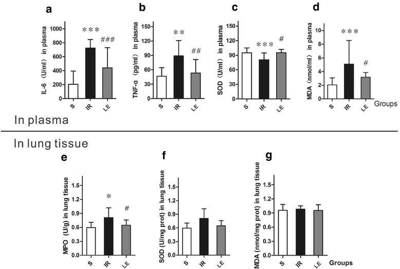
Fig. 3.

Concentration of IL-6, TNF-α, SOD activity and MDA in plasma and MPO, SOD activity and DA levels in lung tissues(n = 8 in Each Group). IL-6, interleukin-6; TNF-α, tumor necrosis factor-α; SOD, superoxide dismutase; MDA, malondialdehyde. MPO, Myeloperoxidase. Data were given as mean ± SD. * p < 0.05 (vs. S), ** p < 0.01 (vs. S), *** p < 0.001 (vs. S). # p < 0.05 (vs. IR), ## p < 0.01 (vs. IR), ### p < 0.001 (vs. IR). a Concentration of IL-6 in plasma displayed differences in the three groups (P = 0.001). b Concentration of TNF-α in plasma displayed differences in the three groups (P < 0.001). c Concentration of SOD activity in plasma displayed differences in the three groups (P =. d Concentration of MDA in plasma displayed differences in the three groups (P = 0.009). e Concentration of MPO Activity in lung tissues displayed differences in the three groups (P = 0.028). f Concentration of SOD Activity in lung tissues displayed no differences among the three groups (P = 0.089). g Concentration of MDA in lung tissues displayed no differences among the three groups (P = 0.170)
