
Fig. 5.
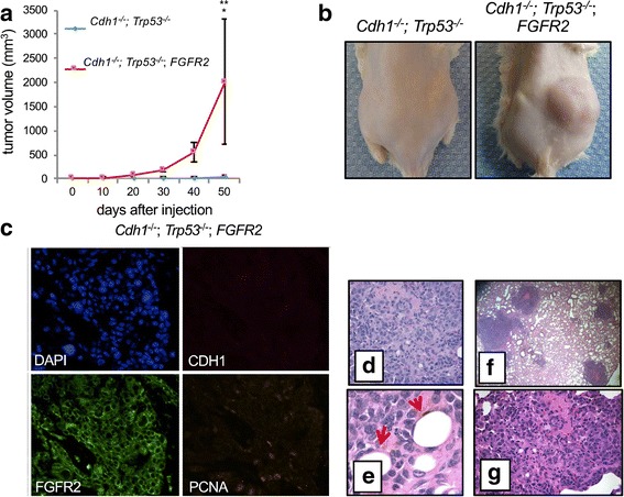
Gastric organoid tumor model. Gastric organoids with the indicated genotypes are shown. a Tumor volumes were measured over time post-injection. Gastric organoids were dissociated and subcutaneously injected into the flanks of NOG mice. Cdh1 -/-;Trp53 -/- is shown in blue, and Cdh1 -/-;Trp53 -/-;FGFR2 is shown in red. Error bars represent SEM, and asterisks indicate p < 0.04. b Images indicate tumor growth at 50 days post-injection. c Overexpression of FGFR2 was confirmed in the tumor derived from Cdh1 -/-;Trp53 -/-;FGFR2 organoids. d–e Histological analysis of the Cdh1-/-;Trp53-/-;FGFR2 tumors confirms the presence of poorly differentiated adenocarcinoma with signet ring as indicated by arrows. f, g After flank injections with dissociated organoids, histological analysis of murine lungs after 50 days revealed metastatic gastric adenocarcinoma with signet ring features at low (f) and high (g) magnification