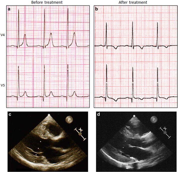
Figure 1.
Electrocardiogram (ECG) and trans-thoracic echocardiography (TTE) before (left) and after (right) treatment with carnitine supplementation. (a) Pretreatment precordial ECG tracings of V4 showing tall peaked T-waves, and high voltage R-wave and a short QTc interval (318 ms). (b) Precordial ECG tracing (lead V4) after 3 months of treatment showing the decreased T-wave amplitude, inversion of the T-wave and QTc interval normalization (400 ms). (c) At pretreatment TTE, LVPWd and the IVSd measured 8.3 and 10.5 mm, respectively. (d) After 3 months of the treatment, both LVPWd and IVSd decreased and measured 6.8 and 7.9 mm, respectively. IVSd, end-diastolic interventricular septal thickness; LVPWd, end-diastolic left ventricular posterior wall thickness.

