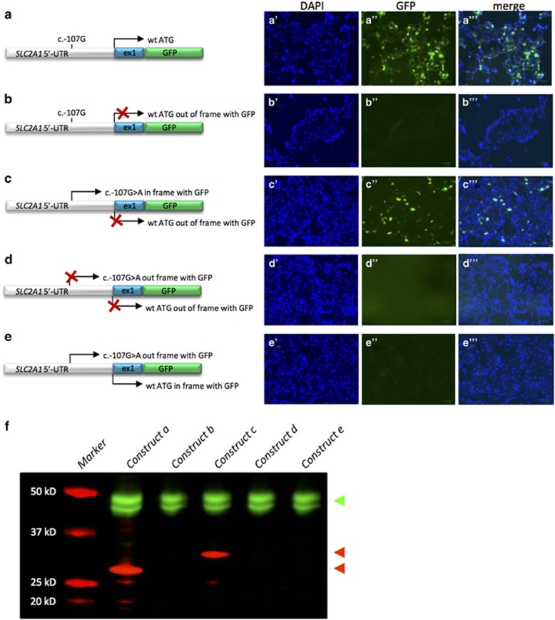
Figure 2.
Functional characterization of SLC2A1 c.-107G>A. HEK293T cells were transfected with constructs of the 5′-UTR and exon 1 of SLC2A1 fused to GFP. Left: cartoons of the constructs used. The positions and possible use (arrow) of the wild-type initiation codon (a–e) and the novel initiation codon introduced by the c.-107G>A variant (c–e) are indicated. If no expression is anticipated, the arrow is crossed out (X). Right: representative images of cells transfected with the respective constructs with signals for nuclei by DAPI staining (blue) or green fluorescent protein (green). (f) Western blot of the SLC2A1-GFP fusion proteins created by the constructs in HEK293T cells. An antibody directed against tubulin was used as control (in green, top arrow head), whereas an antibody against GFP was used to detect the SLC2A1-GFP fusion (in red). The fusion proteins resulting from usage of the novel initiation site or the wild-type initiation codon are 32 kD (upper red arrow head) and 29 kD (lower red arrow head) in size, respectively.

