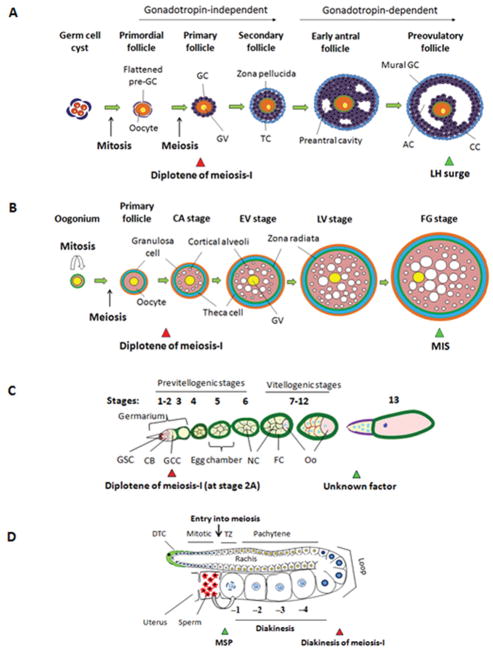
Figure 3.
Stages of oocyte development and meiotic resumption. (a) Formation of primordial follicles varies by species. Primordial follicles develop in utero in humans, versus 1–2 days after birth in mice. A primary follicle, developed from the primordial follicle, consists of a prophase-I–arrested primary oocyte surrounded by somatic granulosa cells (GC). By the secondary stage, the primary oocytes grow, granulosa cells proliferate, and an additional layer of somatic thecal cells (TC) forms outside the basement membrane of the follicle. In both mice and humans, preantral follicle development does not require pituitary gonadotropins. At puberty, FSH secreted by the anterior pituitary promotes further granulosa cell proliferation and survival. As a fluid-filled antrum cavity (AC) begins to form, secondary follicles become early antral follicles. The full-grown primary oocyte is surrounded by proximal cumulus cells (CC) and distal mural granulosa cells in preovulatory follicles. After an LH surge, oocytes undergo meiotic maturation. (b) In fish, the oogonium proliferates and generates primary follicles, at which point the oocytes enter meiosis and arrest at diplotene. The oocytes then begin to enlarge, and form follicles. Next cortical alveoli accumulate within the oocytes, followed by vitellogenesis, which results in increase in the size of the oocyte. Full-grown (FG) follicles initiate oocyte maturation. The germinal vesicle migrates from the center of the oocyte to the periphery, and breaks down in response to maturation-inducing steroid (MIS). (c) In Drosophila, the ovariole is composed of the germarium in the anterior-most part followed by a row of progressively older egg chambers. Within region 1 of the germarium, a cystoblast (CB), derived from a germ-line stem cell, divides four times via mitosis to form a 16-cell germ-line cyst (GCC). Meiosis begins at stage 2A of the germarium, and the oocyte (Oo) arrests at diplotene of prophase-I. The posterior-most germ line cell becomes the oocyte, whereas the remaining 15 cells become nurse cells (NC). The egg chamber is surrounded by a single layer of follicular cells (FC). At stage 13, after an unknown developmental or hormonal signal, the oocyte resumes meiosis and progresses to MI. (d) In C. elegans, adult hermaphrodites have two U-shaped gonads containing germ cells arranged in a distal-to-proximal polarity with respect to the somatic distal tip cell (DTC) at the distal end and the proximal spermatheca/uterus. Germ cells proliferate mitotically at the distal end, known as the “mitotic zone”. At the proximal end of the mitotic region, germ cells switch to meiotic prophase, where they are first in leptotene/zygotene (transition zone [TZ]) and then progress through an extended pachytene followed by diplotene and diakinesis around the loop region. Primary arrest of C. elegans oocytes occurs at diakinesis. In response to sperm and its secreted factor, MSP, the most proximal oocyte (−1) is induced to undergo meiotic maturation. The red and green triangles indicate prophase-I arrest and meiotic resumption, respectively.

