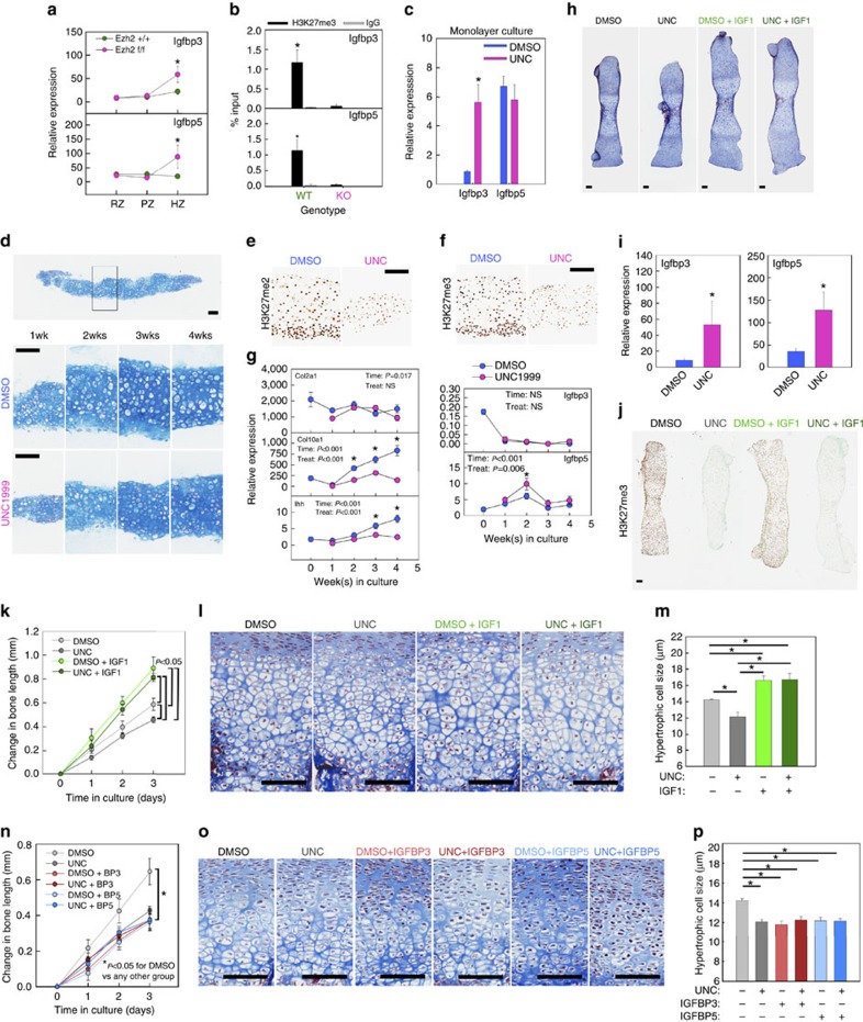
Figure 4. Igfbp3/5 upregulation contributed to defects in chondrocyte hypertrophy.
(a) Relative expression of Igfbp3 and 5 in different zones (RZ, PZ and HZ) isolated from proximal tibial growth plates of 3-day-old Ezh1/2 or wild-type mice. Tissue was isolated by laser capture microdissection, and messenger RNA levels were measured by real-time PCR; *P<0.05 between Ezh1/2 or wild-type mice within the same zone (N=6). (b) ChIP with H3K27me3 antibody (or IgG), followed by real-time PCR to compare levels of H3K27me3 near transcription start site of Igfbp3 and Igfbp5, in chondrocytes isolated from 1-week-old Ezh1/2 or wild-type mice. *P<0.05 between Ezh1/2 or wild-type mice for H3K27me3 (N=6). (c) Relative expression of Igfbp3 and Igfbp5 in monolayer primary chondrocytes isolated from 1-week-old wild-type mice treated with an Ezh1/2 inhibitor (UNC) or vehicle (DMSO). *P<0.05 (N=6). (d) Top panel: Alcian blue-stained histological section of a chondrocyte pellet cultured for 1 week. Middle and bottom panels: higher magnification of DMSO-treated pellets (middle panel) or UNC-treated pellets (bottom panel) at different time points. (e,f) Histological sections of pellet treated with DMSO or UNC for 1 week, immunostained (brown colour) for H3K27me2 (e) or H3K27me3 (f). (g) Relative expression of Col2a1, Col10a1, Ihh, Igfbp3 and Igfbp5 in chondrocyte pellets treated with DMSO or UNC at different time points. P values, two-way analysis of variance for the effect of time in culture and UNC treatment. *P<0.05 between DMSO and UNC treated at a particular time point (N=6). (h) Histological sections of cultured fetal mouse metatarsal bones treated with DMSO or UNC, with or without IGF-I. (i) Relative expression of Igfbp3 and Igfbp5 in metatarsal whole growth plate from h. *P<0.05. (j) Immunohistochemistry (brown colour) for H3K27me3 of metatarsal bones from h. (k–m) Change in length (k) histological sections (l), and quantitative measurements of hypertrophic cell size (m) in fetal metatarsal bones treated with DMSO or UNC, with or without Igf1. Statistical comparison was performed on bone length between different treatment groups at the end of treatment (m). (n–p) Similar to k–m, except for the treatment with Igfbp3/5 instead of IGF-I. *P<0.05, N=6 (m,p). *P<0.05, DMSO versus all other groups (n). Scale bars, 100 μm.

