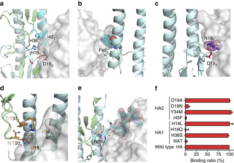
Figure 5. Residues in the natural variants that may affect 3E1 recognition.
(a) Locations of the key residues of HA (shown with side chains) that may affect 3E1 recognition. The HA is shown in cartoon and the 3E1 Fab in surface representation. The colour coding of HA is the same as Fig. 3b. (b) The I45F variant (shown with side chain and dots) in HAs of the H2 subtype viruses could cause steric hindrance with the HCDR3 loop of 3E1 in the HA of Jap57 (H2) structure (PDB code: 2WRD). (c) The D19N variant in HAs of the H13 subtype viruses adopts a different side-chain conformation in the HA of ML77 (H13) structure (coloured in magenta, PDB code: 4KPQ), which could cause steric hindrance with 3E1. (d) The H18Q variant in HAs of the H9 subtype viruses makes relatively weaker interactions with Asn20, Thr37 and His38 in the HA of KO98 (H9) structure (coloured in orange, PDB code: 1JSD), which might interfere with the recognition by 3E1. (e) The glycans at Asn38 in the HAs of the H3 and H7 subtype viruses might obscure the recognition by 3E1. The glycans at Asn38 in the HA of HK68 (H3) structure (PDB code: 4WE4) are shown as cyan sticks. (f) Relative binding ratio of the mutants of WA11 HA with the 3E1 Fab. Bars represent mean±s.d. Data represent a representative experiment from three independent experiments.

