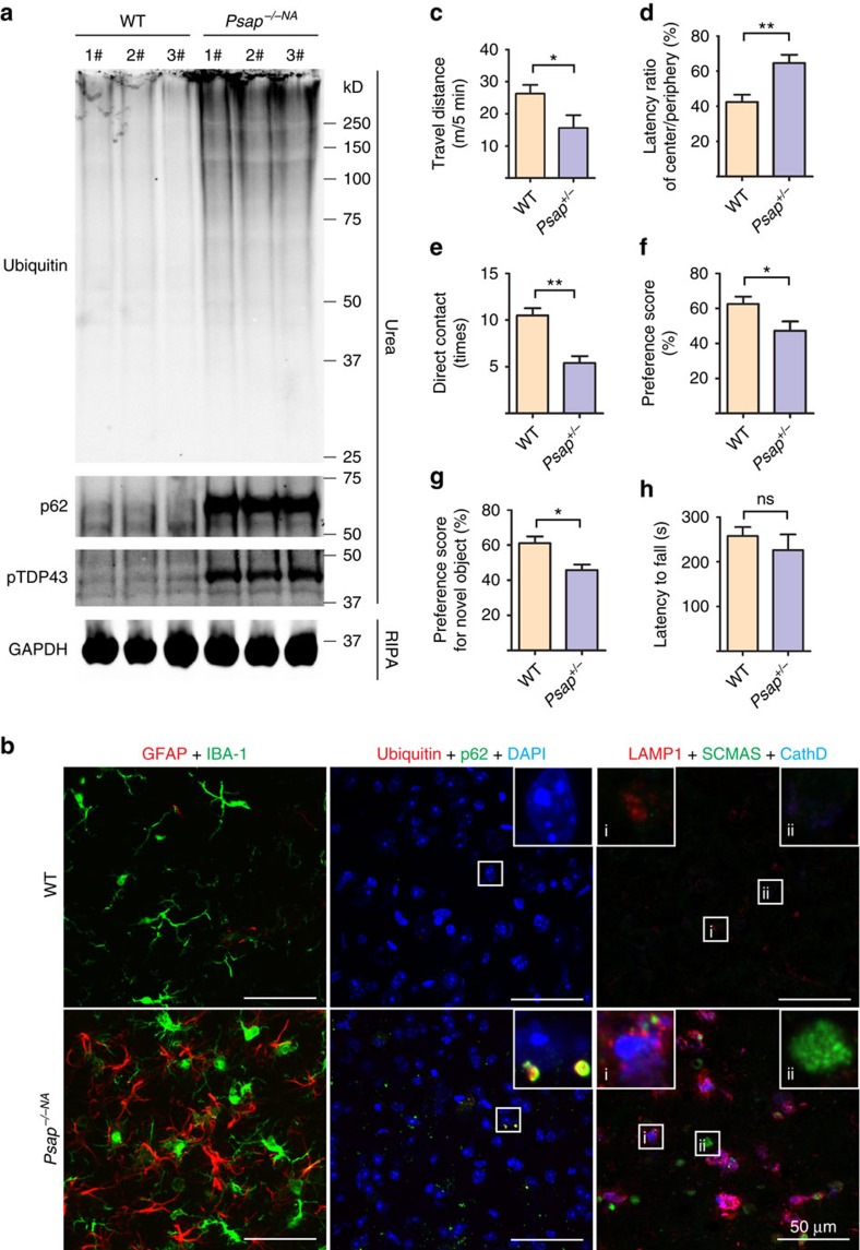
Figure 9. Partial loss of PSAP leads to FTLD-like phenotypes in mice.
(a) RIPA- and urea-soluble fractions from 6-month-old WT and Psap−/−NA brain were blotted with anti-ubiquitin, p62, phospho409/410 TDP-43 and GAPDH antibodies as indicated. (b) Brain sections from 6-month-old WT and Psap−/−NA mice were stained with anti-GFAP, anti-IBA1, anti-ubiquitin, anti-p62 or anti-SCMAS, anti-LAMP1 and anti-CathD antibodies as indicated. Scale bar, 50 μm. A representative glia cell with high LAMP1 levels is shown in inset i and a representative neuron is shown in inset ii for anti-SCMAS staining. The representative images from three mouse brains were shown. (c,d) Twelve-month-old WT and Psap+/− mice were subject to open-field test and Psap+/− mice shows significant reduction in travel distance and increase in latency in the centre arena. (e,f) Psap+/− mice show deficits in the sociability test. Data are presented as mean±SEM. (g) Psap+/− mice show deficits in the novel object test. (h) Psap+/− mice do not show any significant motor deficits in the rotarod test. For all the behavioural tests, WT, n=7; Psap+/−, n=6. *P<0.05; **P<0.01; NS, not significant.

