
Figure 1.
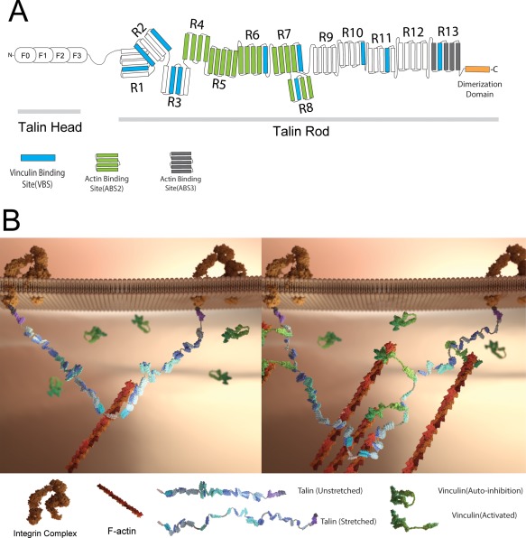
(A) Illustration of the domain structure of full‐length talin. The talin head domain contains a FERM domain (50 kDa), followed by a flexible “neck” (10 kDa) which connects the head domain to its C‐terminal rod domain (220 kDa). The rod domain contains 11 cryptic VBS (drawn in blue). The dimerization domain is a single helix that sits at the end of the rod. (B) Schematics of the talin structure and interaction of the talin dimer with vinculin in cells. A. Illustration of the domain structure of full‐length talin. Talin head domain contains a FERM domain (50 kDa), followed by a flexible “neck” (10 kDa), which connects the head domain to its C‐terminal rod domain (220 kDa). The rod domain contains 11 cryptic VBS (drawn in blue). The dimerization domain is a single helix that sits at the end of the rod domain. B. (left) In the initial stage of FA formation, the talin dimer binds to actin and integrin. At this stage, the cryptic VBSs remain buried among the α‐helical bundles. (right) As the actin filament starts to pull on talin, the formerly buried VBS are revealed to allow vinculin binding, and cause more actin filament recruitment.