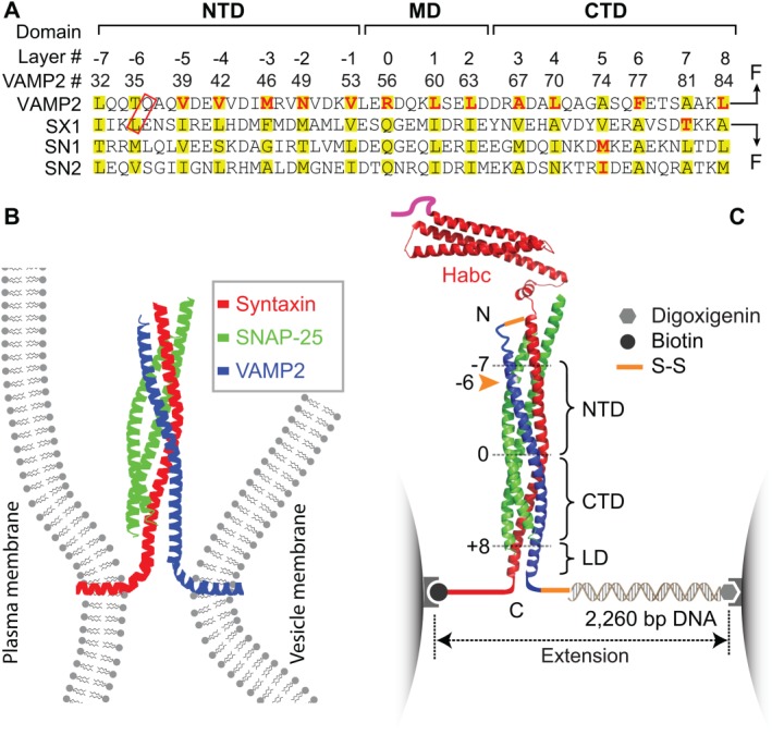
Figure 1.

SNARE domain structures, membrane fusion, and the experimental setup to study SNARE assembly. (A) Amino acid sequences and their domain structures of the SNARE motifs in synaptic syntaxin 1 (SX1), VAMP2, and SNAP‐25B (SN1 and SN2). The amino acids in the hydrophobic layers or the central ionic layer are highlighted in yellow. The amino acids colored red are mutated to test their effects on SNARE assembly. Syntaxin and VAMP2 are cross‐linked at their N‐termini, with one of the cross‐linking sites marked by a red rectangle, and pulled from their C‐termini. The extremely fast middle domain (MD) transition and its associated state 3 can only be resolved in some experiments and is often mixed with the CTD transition, as is shown in C. (B) SNARE proteins couple their folding and assembly to draw two membrane into proximity and force them to fuse. An assembled synaptic SNARE complex is depicted to bridge the plasma membrane and the vesicle membrane. (C) Experimental setup to pull a single cytoplasmic domain of the synaptic SNARE complex using optical tweezers (OTs). The assembled SNARE complex consists of an N‐terminal Habc domain in a antiparallel three‐helix bundle, the core four‐helix bundle domain, and a linker domain (LD) in a two‐stranded coiled coil. The four‐helix bundle contains an N‐terminal domain (NTD) and a C‐terminal domain (CTD) that are separated by a central ionic layer (“0” layer). Adapted from Ma et al.53
