
Figure 1.
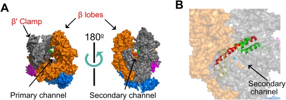
High‐resolution structure of Thermus aquaticus core RNAP (PDB 1HQM). Figure generated using PyMol. (A) The five RNAP subunits are represented with different colors: The two α subunits are blue, β′ is grey, β is orange, and ω is magenta. Two orientations (related by ∼180° rotation) are shown, with the LEFT showing the trailing edge of RNAP facing upstream DNA and the RIGHT showing the leading edge facing downstream DNA. The cleft between the β′ clamp and β lobes (the pincers) forms the primary channel, while the secondary channel is arranged on the opposite face of RNAP. (B) The catalytic magnesium (cyan sphere), bridge helix (red), trigger loop (green), and F‐loop (yellow) of the active site can be seen from the downstream facing side of RNAP.