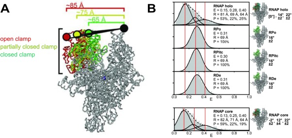
Figure 3.

RNAP clamp conformation during different steps of transcription. Reprinted from Chakraborty et al.49 with permission. (A) The labeling positions of the FRET dye pair at the tip of the RNAP pincers (Cy3B donor on β′ clamp tip, Alexa647N acceptor on β lobe tip) distinguishes three clamp conformations (distances shown) during different steps of transcription: the closed clamp‐higher FRET configuration (green), the partially closed clamp‐medium FRET configuration (yellow), and the open clamp‐lower FRET configuration (red). (B) FRET histograms from single molecule FRET experiments. Three vertical lines mark the mean efficiency of FRET transfer “E” for each of the three clamp conformations. The mean distance calculated from the mean efficiency is shown as is the percentage of each subpopulation.
