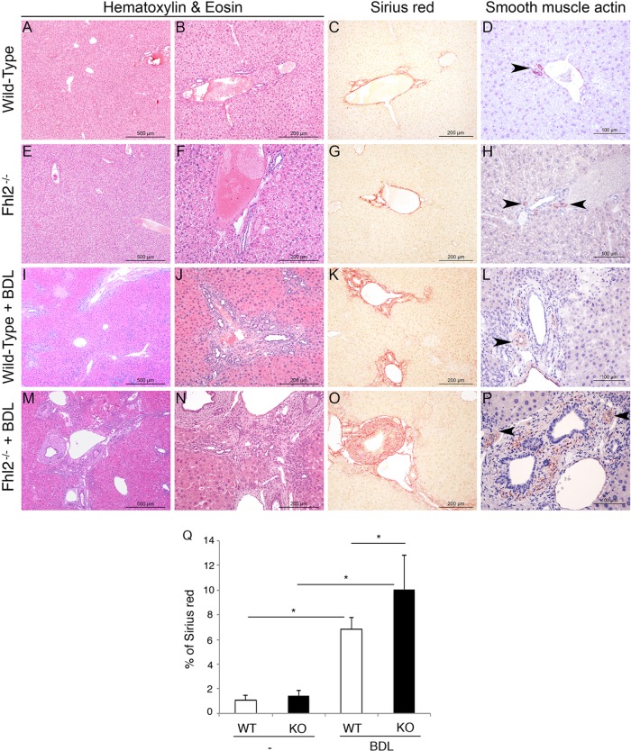
FIG 5.
Enhanced hepatic fibrosis in Fhl2−/− mice. Fhl2−/− mice (n = 7) and WT mice (n = 6) underwent BDL. H&E staining of liver parenchyma was performed with control WT and Fhl2−/− mice (A, B, E, and F) or mice subjected to BDL (I, J, M, and N). Hepatic fibrosis was assessed by Sirius red staining (C, G, K, and O). Quantification is shown in panel Q. Expression of α-SMA was determined by immunohistochemistry (D, H, L, and P). The data presented are the means and SD obtained from at least five mice in each group. *, P < 0.05.

