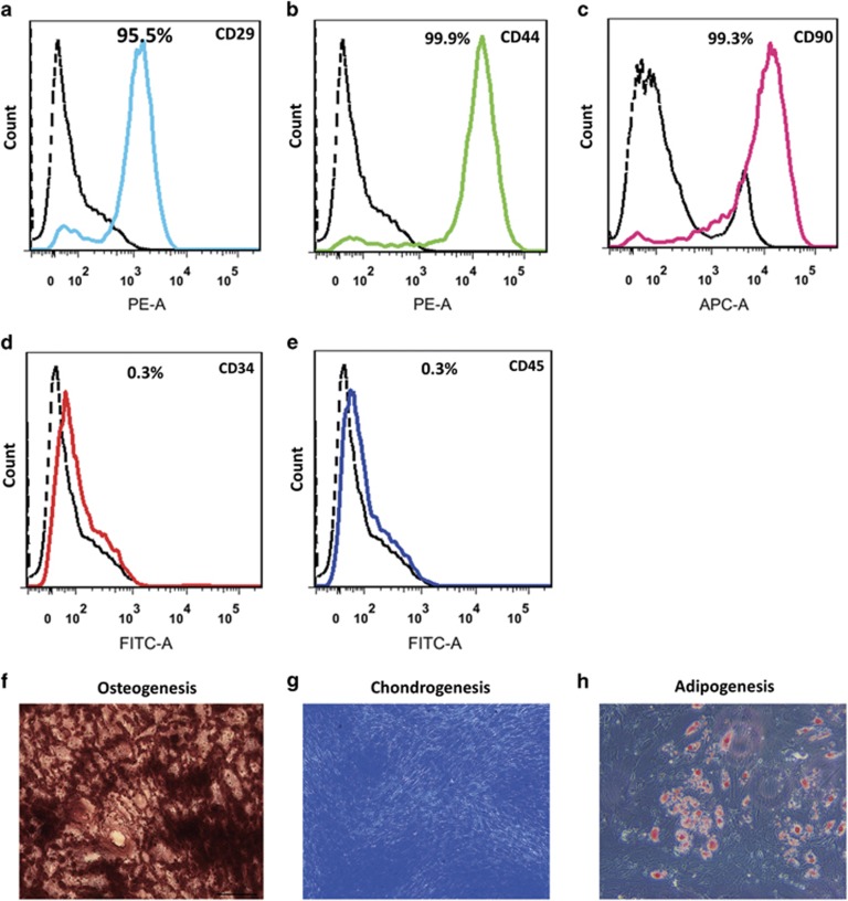
Figure 1.
Characterization of hMSCs. Nearly all of the cells acquired expressed the cell surface markers PE-CD29 (a), PE-CD44 (b), APC-CD90 (c), and negative for the endothelial cell surface marker FITC-CD34 (d) and hematopoietic surface marker FITC-CD45 (e). The osteogenesis, chondrogenesis and adipogenesis differentiation of hMSCs was induced and visualized by alizarin red staining (f, dark red), toluidine blue staining (g, dark blue), and oil red O staining (h, red), respectively

