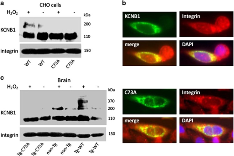
Figure 1.
Integrins coimmunoprecipitate with KCNB1 channels. (a) CHO cells were transfected with cDNA encoding human WT or C73A HA-tagged KCNB1 subunits and lysed in control conditions or subjected to an oxidative insult (1.0 mM H2O2 for 5 min), prior to lysis. In the upper immunoblot, integrin-α5 immunoprecipitates were visualized with anti-HA antibodies to detect KCNB1 protein. Total lysates (lower blot) were stained with anti-integrin-α5. Integrins were detected in a single ∼150 kDa band. (b) Representative images of CHO cells demonstrating colocalization of WT and C73A with integrins. The cells were transfected with pEGFP-N1-WT or pEGFP-N1-C73A and stained with integrin primary antibody and rhodamine-conjugated secondary antibody. Images were analyzed using the ImageJ software. (c) Brain lysates from the indicated genotypes were treated in control conditions or in the presence of 1.0 mM H2O2 for 5 min, prior to the addition of sample buffer. In the representative immunoblots, integrin-α5 immunoprecipitates were visualized with an antibody against mouse and human KCNB1. Total lysates were stained with anti-integrin-α5

