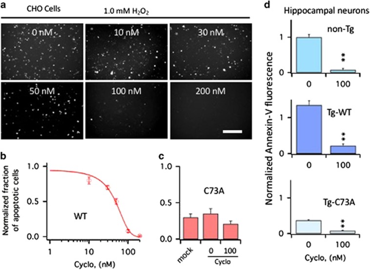
Figure 2.
Cyclo protects CHO cells and primary hippocampal neurons from apoptosis induced by oxidation of KCNB1. (a) Representative examples of Annexin-V staining in CHO cells transfected with WT. The cells were exposed to 1.0 mM H2O2 for 5 min and then incubated 6 h in the presence of Cyclo at the indicated nanomolar concentrations. At the end of the 6-h incubation period, cells were stained with Annexin-V and photographed. Size bar 100 μm. (b) Number of WT-transfected CHO cells subjected to an oxidative challenge positive to Annexin-V in the absence/presence of varying concentrations of Cyclo. Data are normalized to the number of cells positive to Annexin-V in the absence of Cyclo. Data were fit to a sigmoidal function (equation (1)) with an IC50=41.4 nM. N=3 experiments. (c) Number of mock or C73A-transfected CHO cells positive to Annexin-V subjected to an oxidative challenge in the absence or presence of 100 nM Cyclo. N=3 experiments. (d) Quantitative assessment of Annexin-V fluorescence (proportional to the number of cells undergoing apoptosis) in primary hippocampal neurons of the indicated genotypes subjected to an oxidative challenge. After wash out, cultures were incubated in the absence/presence of 100 nM Cyclo for 6 h and then stained with Annexin-V. Fluorescence was assessed using the ImageJ software and data are normalized to non-Tg neurons in the absence of Cyclo. P=1.8 × 10⁻¹⁰ (one-way analysis of variance), **P<0.01 (Tukey's post hoc). N=3 embryos/genotype

