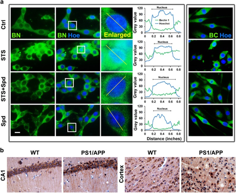
Figure 7.
Nuclear translocation of Beclin 1 in damaged neurons. (a) Cells were treated with STS, STS+Spd or Spd. After incubation, cells were stained with anti-Beclin 1 N terminal (BN) or C-terminal (BC) antibody. The nuclei were counterstained with Hoechst 33342. Scale bar, 10 μm. Curves indicated the colocalization between Beclin 1 (green) and Hoechst (blue) and correlated to the lines drawn in the enlarged images. The x axis represented the distance (inches) along the line and the y axis indicated the pixel intensity. (b) Hippocampal CA1 and cortical areas were obtained from 12-month-old PS1/APP (AD) mice (n=6) and age-matched WT control (n=6). Brain slides were immunostained with anti-Beclin 1 N terminal antibody and the nuclei were counterstained with hematoxylin. Scale bar, 20 μm

