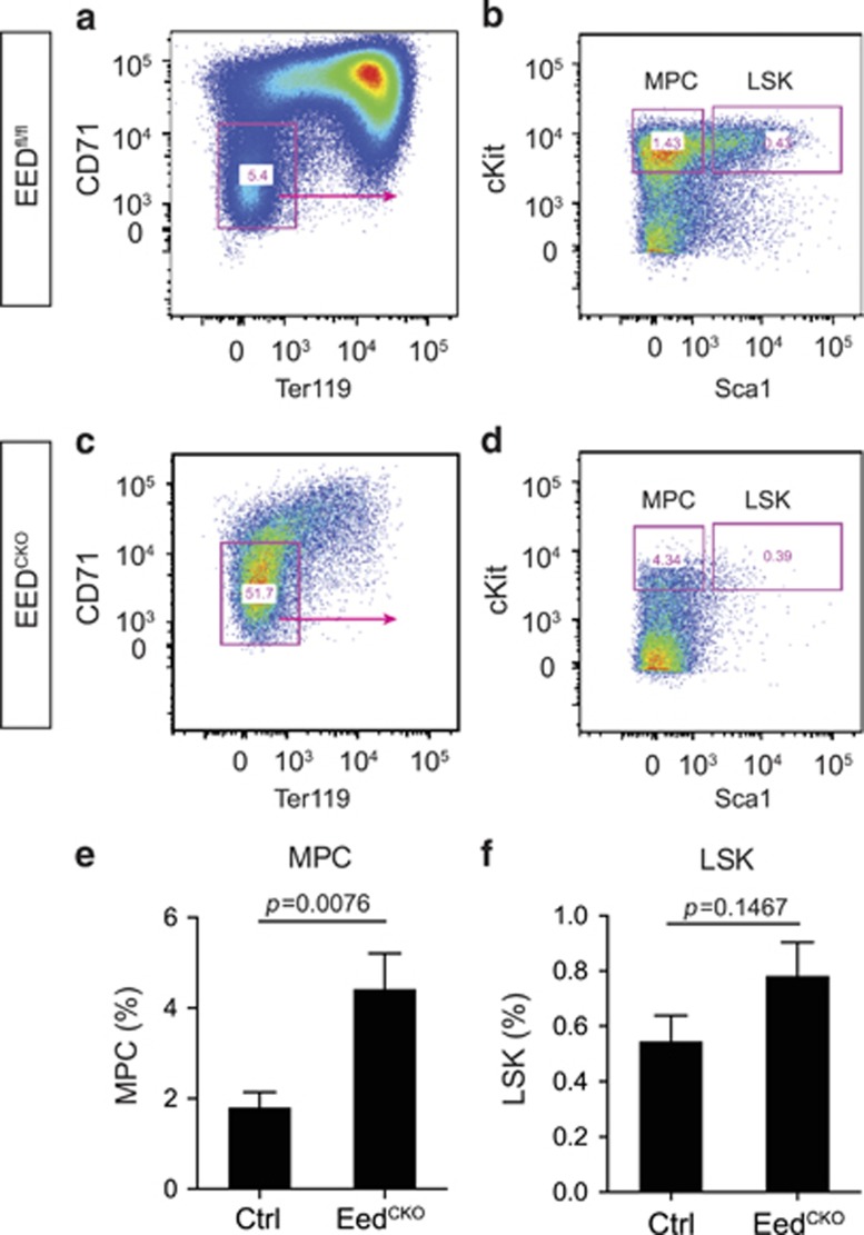
Figure 5.
Eed deletion resulted in defective development of myeloid progenitor cells. (a–d) Flow cytometry analysis of myeloid progenitor cells (MPCs) and LSK cells from control and EEDCKO fetal liver. (e) Quantitative analysis of MPCs. Unpaired t-test, n=3–6. (f) Quantitative analysis of LSK cells. Unpaired t-test, n=3–6

