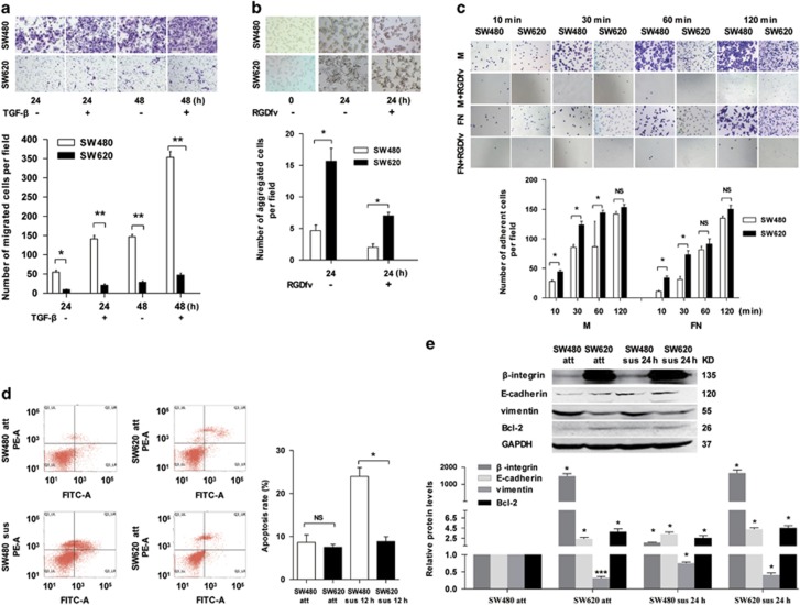
Figure 1.
SW480 cells exhibit stronger migration activity but weaker cell adhesion and anoikis resistance than SW620 cells. (a) Transwell migration assay of SW480 and SW620 cells were seeded and cultured in the Boyden chamber well with or without TGF-β for 24 and 48 h. Above: representative images. Below: quantitative results of three independent experiments (*P<0.05, **P<0.01). (b) Cell–cell adhesion assay of SW480 and SW620 cells suspension cultured with or without cell adhesion inhibitor (RGDfv) for 24 h. Above: representative images, the photomicrographs were taken at × 100 magnification. Below: quantitative results of three independent experiments (*P<0.05). (c) Cell–matrix adhesion assay of SW480 and SW620 cells adhering to FN and Matrigel (M) with or without RGDfv for 10, 30, 60 and 120 min, respectively. Above: representative images (100 × ). Below: quantitative results of three independent experiments. The adherent cells were measured by software of ImageJ (NS indicates no statistical difference between the groups, *P<0.05). (d) FACS analysis of the apoptosis of attached/suspended (att/sus) SW480 and att/sus SW620 cells cultured for 12 h. Left: representative images. Right: quantitative results of three independent experiments (NS indicates no statistical difference between the groups, *P<0.05). (e) Western blot results showing E-cadherin, vimentin, β-integrin and Bcl-2 expression in SW480 and SW620 cells with attached (att) and suspended (sus) growth manner. The expression levels were normalized to GAPDH (*P<0.05, ***P<0.001)

