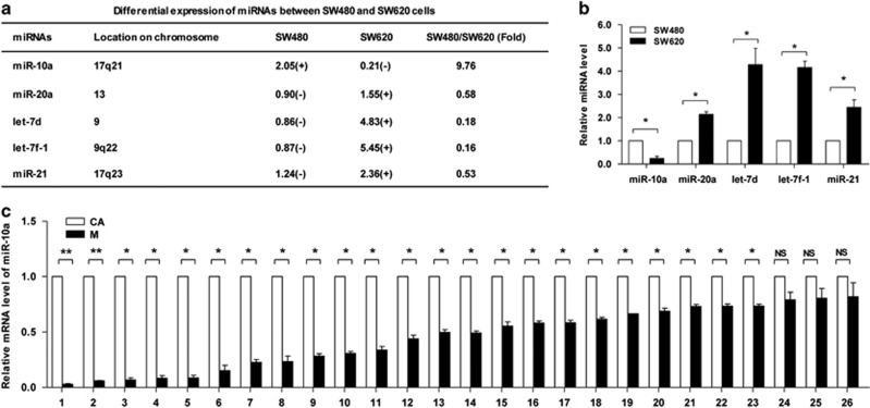
Figure 2.
miR-10a is overexpressed in primary CRC cells and tissues. (a) The miRNAs differentially expressed between SW480 and SW620 cells were analyzed using a miRNA microarray. The five miRNAs exhibiting the significant differences are shown. The miRNAs with a signal background ratio of 1.5 or greater were considered to represent positive expression (+), and the others were considered to represent negative expression (−). (b) RT-qPCR results of the five miRNAs in SW480 and SW620 cells (*P<0.05). (c) RT-qPCR results of miR-10a in human primary CRC tissues (CA) and paired lymph node metastases (M). The expression levels were normalized to U6 snRNA (*P<0.05, **P<0.01, NS, not significant)

