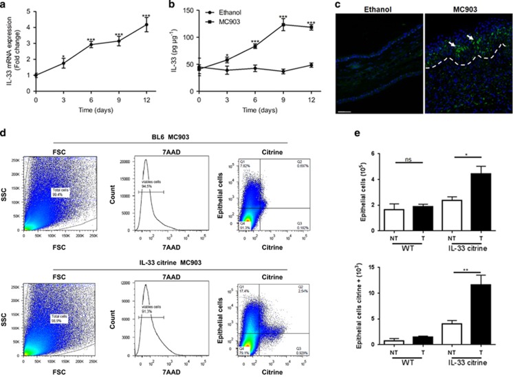
Figure 2.
IL-33 is mainly expressed by epithelial cells and CD11b+ cells. (a and b) Quantification of IL-33 mRNA (a) and protein (b) expression in the BL6 mouse ears for different times. (c) Immunofluorescence analysis of IL-33 in mouse ear skin after 12 days of MC903 treatment. This image is representative of five mice with abscess. Scale bar represents 50 μm. (d and e) Expression of total and citrine Epcam+ cells assessed by flow cytometry of MC903 administrated BL6 and IL-33 citrine reporter mice at day 12. *P<0.05, **P<0.01,***P<0.001. P-values were analyzed by one-way ANOVA in a and e, two-way ANOVA in b. All data are representative of two independent experiments with n=3–5 per group and are means±S.E.M.

