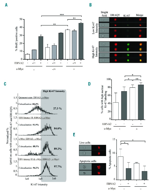
Figure 2.

Proliferation and apoptosis of P493.6 cells after induction of c-Myc programs in EBV-latency III B cells. (A) Percentage of BrdU positive cells was assessed by flow cytometry on P493.6 cells treated with two doses of estradiol (0.1 μM: EBNA2−/+ and 1 μM: EBNA2+) or not (EBNA2−) and/or with tetracycline (100 μg/ml: c-Myc−; 3 μg/ml: c-Myc−/+; and not: c-Myc+) for 48 hours. Data are shown as mean ± standard deviation (n=3 independent experiments). (B to E) P493.6 cells were treated or not with 1 μM estradiol and/or 100 μg/ml tetracycline for 48 hours to induce either cell quiescence (EBNA2−, c-Myc−), EBV-latency III alone (EBV+, c-Myc−), c-Myc alone (EBNA2−, c-Myc+), and both c-Myc and EBV-latency III (EBNA2+, c-Myc+). (B,C and D) Percentage of cells with strong staining of the Ki-67 proliferation marker in the nucleus, assessed by imaging flow cytometry. (B) Examples of cells under the c-Myc program with low or high Ki-67 expression. Selection of cells was based on colocalization of Ki-67 and the DRAQ5® nuclear dye. (C) Representative monoparametric histograms of Ki-67 intensity gated on cells with colocalization of Ki-67 and DRAQ5®. Each cell condition is indicated at the top left of each histogram. Threshold for strong Ki-67 labeling is indicated by the gray box. Percentage of cells with high Ki-67 labeling is indicated on the top right of each histogram. (D) Total percentage of high nuclear stained Ki-67 cells in the four culture conditions of P493.6 cells. Data are shown as mean ± standard deviation (n=5 independent experiments). (E) Apoptosis of the uncolocalized Ki-67 and DRAQ5® population using imaging flow cytometry. Left panel: example of living and apoptotic cells under the c-Myc condition. Right panel: total percentage of apoptotic cells in the four culture conditions. Quantification was based on morphometric parameters.29 Data are shown as mean ± standard deviation (n=3 independent experiments). Statistical significance was determined by Student’s t-test (*** P<0.001; **P<0.01; *P<0.05). BrdU: 5-bromo-2′-deoxyuridine; EBNA2: Epstein-Barr virus nuclear antigen 2; EBV: Epstein-Barr virus; DRAQ5: deep red anthraquinone 5.
