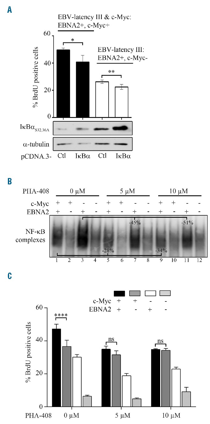
Figure 3.

Activation of NF-κB in P493.6 cells after induction of c-Myc on EBV-latency III. P493.6 cells were treated or not with estradiol 1 μM (EBNA2− or EBNA2+) and/or with tetracycline at 100 ng/mL (c-Myc+ or c-Myc−) for 48 hours. (A) Percentage of BrdU positive cells after transient transfection with the pCDNA.3 empty vector (Ctl) or expressing the super-repressor IκBαS32,36 A for 48 hours (upper panel). Representative analysis of IκBαS32,36A, and α-tubulin expression by Western blot (lower panel). (B and C) Effect of the IKK2 inhibitor PHA-408 (5 μM and 10 μM) for 48 hours on NF-κB. Representative DNA binding by EMSA (panel B) and BrdU incorporation (panel C). In the histograms, data are shown as the mean ± standard deviation (at least 3 independent experiments). Statistical significance was determined by Student’s t-test (****P<0.001; **P<0.01; *P<0.05). ns: not significant; BrdU: 5-bromo-2′-deoxyuridine; EBNA2: Epstein-Barr virus nuclear antigen 2; EBV: Epstein-Barr virus; Ctl: control; NF-κB: nuclear factor-κB.
