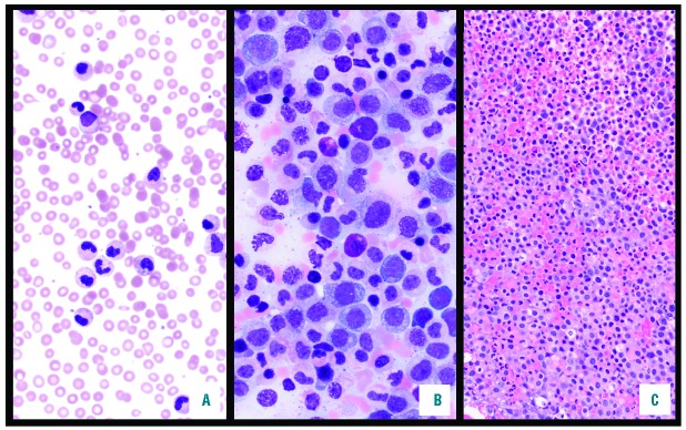
Figure 2.
Peripheral blood smear and bone marrow aspirate and biopsy for the patient at the time of diagnosis. The peripheral smear (A, 600×) is consistent with leukocytosis with a left shift, characterized by a prominent increase in mature neutrophils. This was in accordance with the aspirate smear (B, 600×), which revelated no overt dysplasia or increase in blasts, with mild reticulin fibrosis. The core biopsy specimen (C, 200×) is markedly hypercellular (>95%) for age, and is packed with sheets of left shifted myeloid forms.

