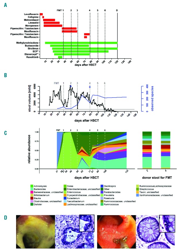
Figure 1.

Characteristics of patient 1. A. Antibiotics (red) and immunosuppressants (green) given to patient 1 before, during and after the introduction of FMTs. ECP, extracorporeal photopheresis. In total, 15 ECP sessions (*) were applied and 13 doses (**) of etanercept 25 mg were given. Methylprednisolone was gradually reduced after FMT 3 (d+58) from 2 mg/kg/d to 1 mg/kg/d at d+111 and maintained beyond discharge. B. Longitudinal illustration of stool volumes [ml/day, black lines, left Y-axis] and richness [observed species, blue lines, right Y-axis] of patient 1 before, during and after six FMTs (grey dashed vertical lines). The horizontal black line represents 500ml of stool volume. Blue crosses represent the bacterial richness (number of observed species) in donor stools at FMT 2 and 4. Stool volumes peaked with 6200 ml at d+47 and substantially decreased to normal values after commencement of FMTs. After FMT 6, diarrhea subsided. C. Fecal microbiota analysis (16S rRNA gene analysis) of patient 1 before, during and after six FMTs (grey dashed vertical lines). Colors represent different taxa on the genus level according to their relative abundance (Taxonomic groups with an abundance less than 2% of the overall abundance are summarized as “Other”). Stool specimens were obtained either before/between FMTs or by aspiration during the colonoscopy before the FMT. Following the first three FMTs, there was no persistent colonization of the introduced stool microbiota (transient colonization after FMT 3). After FMT 4, colonization by donor stool is evident. D. Left panel: Patient 1 before and during FMTs. Endoscopy reveals severe ulcerative ileitis without evidence of any normal ileal mucosa. Histologically severe graft-versus-host disease was confirmed with partial loss of crypts with multiple apoptoses. At the top a completely destroyed crypt (20× magnification). Insert: higher magnification of apoptosis (40×). Right panel: Patient 1 at 37 days after FMT 6. Macroscopically regenerated ileal mucosa with a healed ulcer (center). Histologically mild graft-versus-host disease with a crypt with a single apoptosis on the left (9 o’clock) (20× magnification). Insert: higher magnification of apoptosis (40×).
