
Figure 2. An intersectional strategy to remove B6 cells from the developing retina.
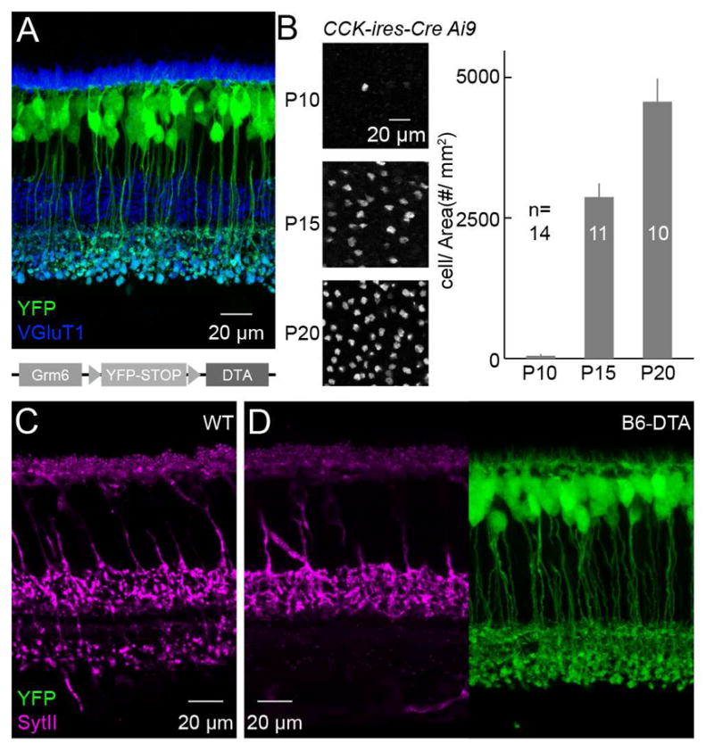
(A) Representative image of a retinal section showing YFP expression in a Grm6L-YFP-DTAcon mouse retina without Cre-recombination. Schematic at the bottom illustrates the genetic construct used to generate Grm6L-YFP-DTAcon mice. (B) Representative images (left) and summary data (right) for the timecourse of tdTomato expression in CCK-ires-Cre Ai9 mice. P10: 48.5 ± 30.5 # / mm2 (mean ± SEM); P15: 2872.2 ± 245.6 # / mm2; P20: 4569.3 ± 411.9 # / mm2. (C and D) Vertical sections of wild-type (C) and B6-DTA (D) retinas stained for SytII and, in (D), YFP. SytII and YFP signals from the same section are shown side by side in (D).