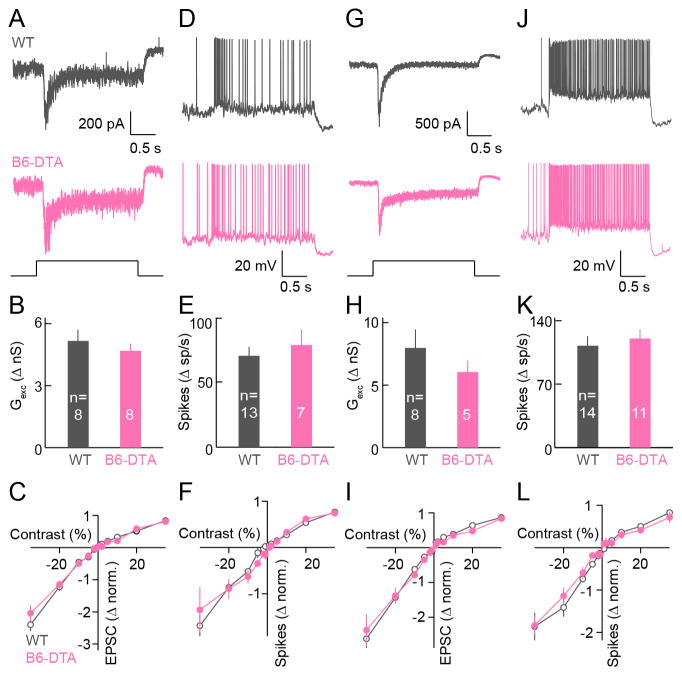
Figure 6. Cell-type-specific rewiring preserves contrast responses of ONα-RGCs in B6-DTA mice.
(A and D) Representative EPSC traces (A) and spike responses (D) of ONα-RGCs in wild-type (grey) and B6-DTA (pink) mice in illumination conditions preferentially activating rods (mean intensity: 1.5 R*). (B and E) Summary data of excitatory conductances (B) and spike responses (E) of ONα-RGCs in wild-type (grey) and B6-DTA (pink) mice in illumination conditions preferentially activating rods. In (B), WT: 5.16 ± 0.55 nS; B6-DTA: 4.69 ± 0.35 nS; p = 0.48. In (E), WT: 70 ± 6.9 sp / s; B6-DTA: 78.3 ± 11.9 sp / s; p = 0.56. (C and F) Contrast response functions of the normalized excitation (C, WT: n = 11; B6-DTA: n = 9) and spike rate (F, WT: n = 12; B6-DTA: n = 10). (G and J) Analogous to A (G) and D (J), but in conditions preferentially activating cones (mean intensity: 1,500 R*). (H and K) Analogous to B (H) and E (K) but in illumination conditions preferentially activating cones. In (H), WT: 7.98 ± 1.48 nS; B6-DTA: 6.05 ± 0.92 nS; p = 0.29. In (K), WT: 112.4 ± 10.5 sp / s; B6-DTA: 120.4 ± 9.9 sp / s; p = 0.59. (I and L) Analogous to C (I, WT: n = 9; B6-DTA: n = 5) and F (L, WT: n = 14; B6-DTA: n = 8) but in illumination conditions preferentially activating cones.

