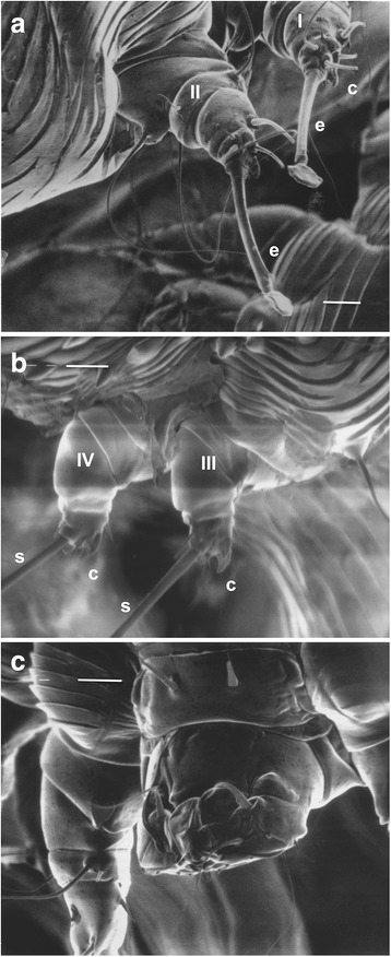
Fig. 2.

Scanning electron micrographs of female Sarcoptes scabiei var. canis. a Legs I and II showing tarsus with claws (c) and stalked empodium (e) that terminates in a pad. b Legs III and IV showing two claws (c) and long seta (s) on the tarsus. c Gnathasoma (pedipalps and chelicerae) and leg I. Scale-bars: a, 10 μm; b, 10 μm; c, 10 μm
