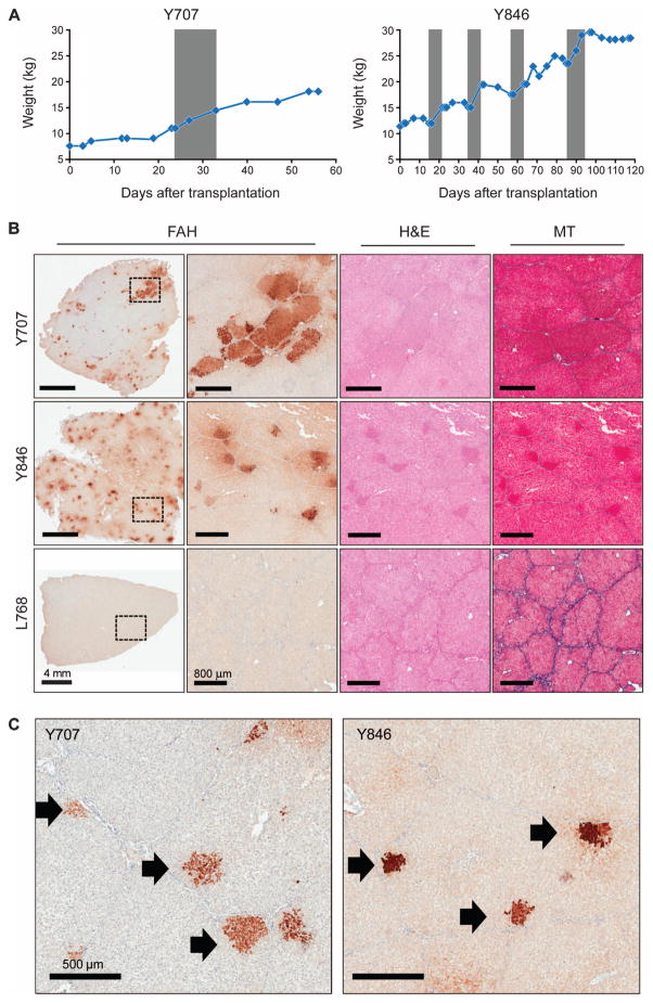
Fig. 3. LV-Fah–transduced hepatocytes proliferate extensively in vivo in pigs.
Animals Y707 and Y846 were euthanized at 2 and 4 months after transplantation, respectively. Control Fah−/− pig L768 did not receive any gene-corrected hepatocytes but was cycled on and off NTBC. (A) Weight stabilization of pigs Y707 and Y846. Gray areas are times on NTBC (for Y707, from day 24 to day 32; for Y846, from days 14 to 21, 35 to 42, 57 to 64, and 86 to 93). (B) Representative liver tissues stained for FAH are shown at low and high magnification for each animal; the dotted rectangle indicates the area of enlargement. Serial sections of H&E–and Masson’s trichrome (MT)–stained liver are also provided. Scale bars, 4 mm (low-magnification FAH); 800 μm (all other images). (C) FAH staining of liver tissue from pigs Y707 and Y846, identifying individual expanding hepatocyte nodules (black arrows).

