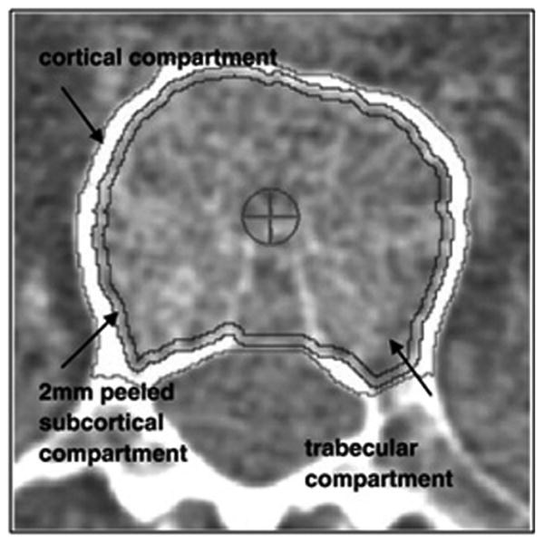
Fig. 1.

Cross-sectional view of the trabecular region of interest in the lumbar spine. Trabecular vBMD included this region only, whereas integral vBMD also included the cortical compartment. Both exclude the posterior elements that DXA measures of BMD incorporate, thus allowing CT measures to more precisely capture BMD of the vertebral body itself. Reprinted with permission from Elsevier from: Engelke K, Mastmeyer A, Bousson V, Fuerst T, Laredo J-D, Kalender WA. Reanalysis precision of 3D quantitative computed tomography (QCT) of the spine. Bone, 2009:44(4):566–72.
