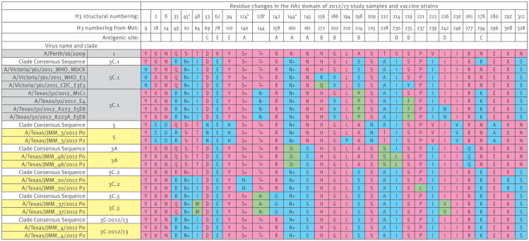
Figure 2. HA amino acid differences across among representative influenza A(H3N2) strains for various HA clades, Texas, 3 November 2012–8 February 2013 (n = 5).
HA: haemagglutinin; Met: methionine; +/- indicates whether the residue is associated with the gain (+) or loss (−) of a potential glycosylation site.
a S45N creates a potential glycosylation site (SSS to NSS), with glycosylation occurring at N45.
b S124N removes a potential glycosylation site (NES to NEN), with glycosylation occurring at N122 (antigenic site A).
c T128N and T128A remove a potential glycosylation site (NWT to NWN or NWA), with glycosylation occurring at N126 (antigenic site A).
d N144D removes a potential glycosylation site (NSS to DSS), with glycosylation occurring at N144 (antigenic site A).
All residue changes that exist in HA among the study samples (yellow), vaccine strains (purple), and clade consensus sequences (white) are provided. All sequences that match the A/Perth/16/2009 strain (clade 1) are coloured in pink, while residues that differ from that strain are shown in blue (first residue difference) or green (second residue difference). Residues associated with the gain (+) or loss (−) of a potential glycosylation site are indicated. Antigenic site assignments are derived from several sources [60-63]. P0 indicates the original clinical swab specimen, while P2 indicates the second-passage viral stock grown in MDCK cells.

