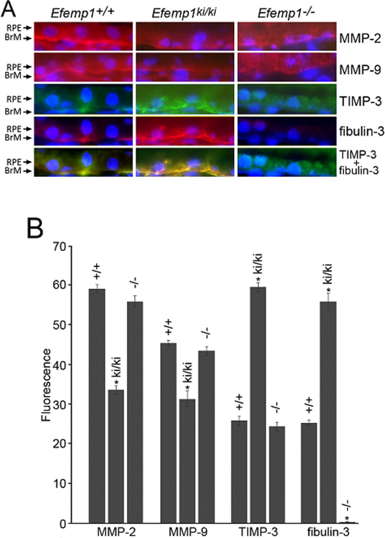
Figure 6.
Immunofluorescence for MMP-2, MMP-9, TIMP-3, and fibulin-3 in the RPE and Bruch's membrane of 9-month-old Efemp1+/+, Efemp1ki/ki, and Efemp1−/− mice. (A) Frozen sections were stained with antibodies (red signal) against MMP-2, MMP-9, TIMP-3 (green signal) or fibulin-3. The nuclei were stained with DAPI (blue signal). Note the more defined linear staining along Bruch's membrane for MMP-2, MMP-9, TIMP-3, and fibulin-3 in Efemp1+/+ and Efemp1ki/ki mice, but a diffuse pattern of staining in RPE cells in Efemp1−/− mice. TIMP-3 and fibulin-3 signals overlapped in the RPE and Bruch's membrane of Efemp1+/+ and Efemp1ki/ki mice. BrM, Bruch's membrane. (B) Immunofluorescence of MMP-2, MMP-9, TIMP-3, and fibuin-3 in the RPE and Bruch's membrane was quantified using Image J software. n = 3 mice per genotype. Error bars indicate the mean ± SD. n = 3 mice per genotype. *P < 0.05 comparing to the values of wild-type controls for each protein in a Student's t-test.

