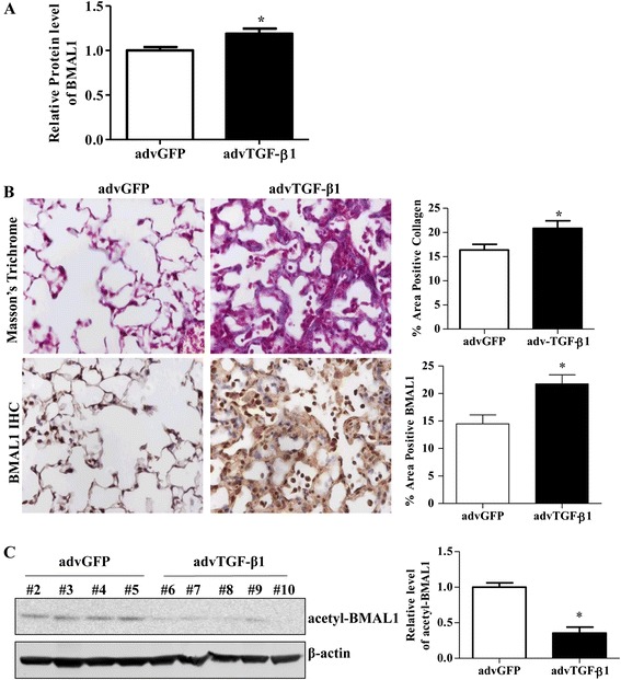
Fig. 2.

Expression of BMAL1 in advTGF-β1-infected mouse lungs. a Densitometric analysis of Western blots of BMAL1 protein expression in the lung homogenate from advTGF-β1-infected mice and Adv-GFP. b Representative images and quantification of Immunohistochemistry analysis to quantify BMAL1 expression and Masson’s trichrome staining to evaluate collagen deposition in lungs from advTGF-β1-infected mice versus Adv-GFP. Positive collagen deposition appears blue. Image J quantification of the respective staining is presented. Data represents the mean ± S.E.M., n > 5, * p < 0.05 vs. advGFP control. c Western blot analysis of acetyl-BMAL1 in the lung homogenate of advTGF-β1-infected mice. Quantification of BMAL1 and acetyl-BMAL1 levels was done by ImageJ and the densities were normalized by β-actin, * p < 0.05 vs. advGFP control
