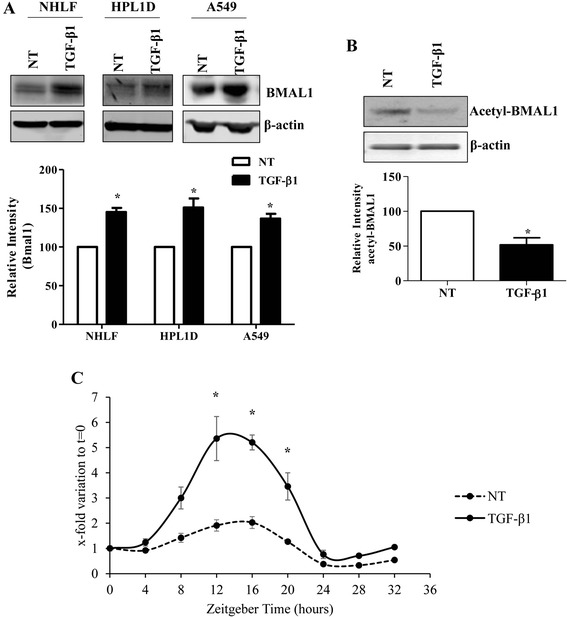
Fig. 3.

Expression of clock gene BMAL1 in response to TGF-β1 in lung cell lines. a Western blot analysis of BMAL1 protein expression in response to TGF-β1 treatment for 24 h in NHLFs, HPL1D, and A549 cells. * p < 0.05 vs. corresponding no treatment group (NT). b HPL1D cells were treated with TGF-β1 at 5 ng/ml for 24 h and Western blot analysis was performed using antibodies against acetyl-BMAL1 (K538). * p < 0.05 vs. NT. c HPL1D cells were treated with TGF-β1 at 5 ng/ml after serum-shock by incubating in media containing 50 % horse serum for 2 h. Cells pellets were harvested every 4 h. Real-time qRT-PCR analysis was employed to investigate the expression level of BMAL1. * p < 0.05 vs. corresponding time points in no treatment (NT) group
