. 2015 Jan 15;97(3):511–520. doi: 10.1189/jlb.1A0714-361R

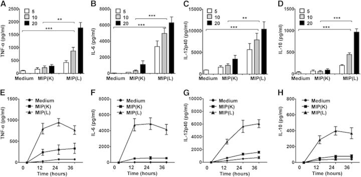
Figure 1. Cytokine production by MIP-stimulated DCs. BMDCs were stimulated with MIP(K) and MIP(L) at MOIs of 5, 10, and 20. Culture supernatants from unstimulated or MIP-stimulated DCs were collected after 24 h and analyzed for TNF-α, IL-6, IL-12p40, and IL-10 by ELISA. Higher levels of these cytokines were produced in response to MIP(L). MIP(K) induced moderate levels of TNF-α, IL-6, IL-12p40, and IL-10 (A–D). For time-kinetics study, BMDCs were stimulated with MIP(K) and MIP(L) at an MOI of 10. Culture supernatants were collected after 12, 24, and 36 h and analyzed for different cytokines. Higher levels of TNF-α, IL-6, IL-12p40, and IL-10 were observed in response to MIP(L) at all time-points (E–H). Mean ± sem of 3 independent experiments is shown. **P < 0.01, and ***P < 0.001.

Figure 1.