. 2015 Jan 15;97(3):511–520. doi: 10.1189/jlb.1A0714-361R

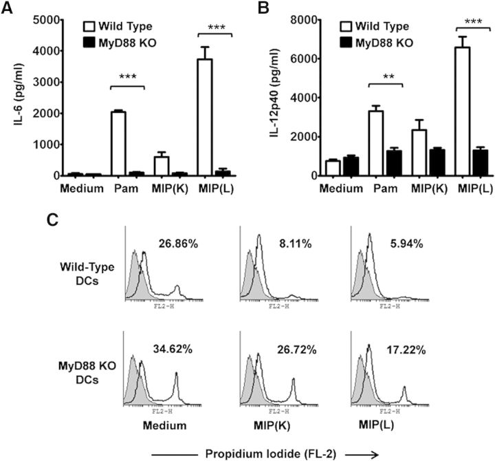
Figure 6. Role of MyD88 in MIP-induced DC activation. DCs derived from MyD88−/− and WT mice were stimulated with MIP(K) or MIP(L) at an MOI of 10, and culture supernatants were analyzed for IL-6 and IL-12p40. Response to MIP(K) and MIP(L) was lost in MyD88−/− DCs, as evident by a drastically reduced production of IL-6 and IL-12p40 by these cells compared with WT cells (A and B). Pam, Pam3CSK4 (TLR2 agonist; 10 ng/ml). Mean ± sem of 3 independent experiments is shown. **P < 0.01, and ***P < 0.001. Role of MyD88 in MIP-induced DC survival was also analyzed. A significant increase in the level of PI-positive cells was observed in MIP(K)-stimulated MyD88−/− DCs compared with MIP(K)-stimulated WT DCs. DC survival efficacy of MIP(L) was also reduced in MyD88−/− DCs compared with WT DCs (C). Representative data of 2 independent experiments are shown.

Figure 6.