. 2015 Jan 12;97(3):583–598. doi: 10.1189/jlb.4A0714-326R

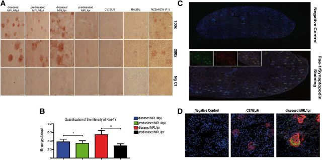
Figure 2. Analysis of Rae-1γ and Mult-1 NKG2D ligands in murine SLE models. (A) Immunohistochemical analysis of Rae-1γ expression in kidney-tissue cryosections from MRL/MpJ (prediseased and diseased), MRL/lpr (prediseased and diseased), C57BL/6, BALB/c, and NZBxNZW(F1) (diseased) mice. Specific staining (red chromogen stains) was observed in the glomeruli of all MRL genotype and diseased NZBxNZW(F1) mice, whereas staining was absent in C57BL/6 and BALB/c controls. Representative images of kidneys from 5 mice/group. (B) Quantification of Rae-1γ staining in MRL mouse glomeruli. Data were analyzed with a 2-tailed, unpaired Student’s t-test (mean ± sd; n = 5/group; **P < 0.0001; *P < 0.0025). (C) Cryosections of diseased MRL/lpr kidney (10 μm) were costained with rabbit anti-mouse synaptopodin (green) and goat anti-mouse Rae-1γ antibody (red), followed by appropriate Alexa-conjugated secondary antibodies and DAPI (blue). All synaptopodin-positive cells corresponded to Rae-1γ-positive cells, indicating glomerulus-specific expression of this NKG2D ligand. (D) Confocal analysis of synaptopodin and Mult-1 expression in C57BL/6 and diseased MRL/lpr kidney sections. Kidney cryosections were stained with rabbit anti-mouse synaptopodin (red) and rat anti-mouse Mult-1 (green), followed by secondary antibodies and DAPI (blue). Mult-1 expression was absent in the C57BL/6 strain.

Figure 2.