. 2015 Jan 12;97(3):583–598. doi: 10.1189/jlb.4A0714-326R

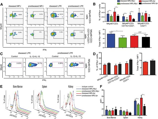
Figure 8. IFN-γ secretion and pSTAT5 of Ficoll-isolated kidney lymphocytes. (A) Ficoll-purified kidney lymphocytes were incubated with PMA, ionomycin, and BFA and stained with anti-NKp46, -CD45, -CD3, -CD11b, -CD27, and -IFN-γ mAb (4 h). Representative flow cytometric density plots show the percentage of gated CD45+NKp46+CD3 and CD45+NKp46+CD3CD11b+CD27 cells positive for IFN-γ. (B) Percentages of CD11bCD27 subsets positive for IFN-γ, and the median fluorescence intensity (MFI) of IFN-γ in NK cells were calculated. (C) As in A, lymphocytes from MRL/lpr mice were stimulated with IL-12 and IL-15 for 6 h, and BFA added in the last 4 h of incubation. Cells were stained as in A. Representative flow cytometry density plots are shown. (D) Percentage of IFN-γ−positive cells in total NKp46+ CD3 and in CD11bCD27 NK subsets and median fluorescence intensity of IFN-γ+ NK cells was calculated. (E) Fresh single-cell suspensions of spleen, BM, and Ficoll-isolated kidney lymphocytes were activated with murine rIL-15 (15 min), fixed, permeabilized, and stained with anti-NKp46, -CD45, -CD3, and -pSTAT5 mAb. Representative flow cytometric histograms show pSTAT5 expression on gated CD45+ NK cells for diseased MRL/MpJ (blue line), prediseased MRL/MpJ (green), diseased MRL/lpr (red), and prediseased MRL/lpr (black) mice. Isotype control staining (gray) is shown in each panel. (F) NK cells were gated for CD45+NKp46+CD3 and the median fluorescence intensity calculated for total pSTAT5 expression in BM, spleen, and kidney. Gates for each population were set by use of unstained or nonspecific mAb isotype controls. Data were analyzed with a 2-tailed, unpaired Student’s t-test (mean ± sd; n = 3–6 mice/group in 3–4 independent experiments; *P < 0.05).

Figure 8.