Fig. 1.

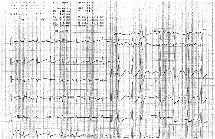
EKG showing diffuse alterations of the repolarization phase; the Q–U prolongation is extreme and the distinction between T wave and U wave is lost, one ventricular extrasystole only is observed, in line with a chronic adaptation.

EKG showing diffuse alterations of the repolarization phase; the Q–U prolongation is extreme and the distinction between T wave and U wave is lost, one ventricular extrasystole only is observed, in line with a chronic adaptation.