Fig. 2.

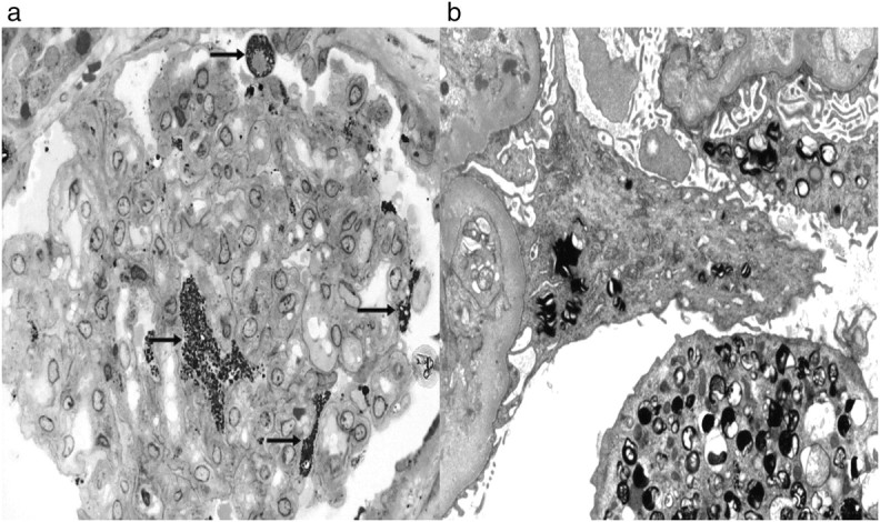
Recipient B—biopsy taken 12 years after transplantation. Lipid deposits (arrows) in podocytes (toluidine blue, ×400) and by electron microscopy (×5200).

Recipient B—biopsy taken 12 years after transplantation. Lipid deposits (arrows) in podocytes (toluidine blue, ×400) and by electron microscopy (×5200).